High-frequency transducers and MFI-HD enhance diagnosis of suspected placental abnormality
By Michael S. Ruma, MD, MPH ∙ Jun 30, 2020 ∙ 3 min read
Placenta Accreta Spectrum (PAS) defines a group of disorders where placental trophoblasts invade into the myometrium of the uterine wall.1 This diagnosis is critical for identification prior to delivery in order to reduce maternal morbidity and mortality. If PAS is present, the placenta will not separate after delivery of the infant and results in substantial hemorrhage, which often requires a hysterectomy to save the life of the mother.
At-a-glance

Obstetric ultrasound is the most important diagnostic tool used to identify this significant abnormality. The ultrasound detection rate of PAS is as high as 80–90%.2 To achieve confidence in the diagnosis, magnetic resonance imaging (MRI) is often used as an adjunctive measure to evaluate suspected placental abnormalities. The detection rate of MRI is no higher than that of ultrasound, ranging 80–90%.1
2D ultrasound is the preeminent modality utilized in the evaluation and prenatal diagnosis of placental abnormalities. The use of color flow Doppler also is utilized to improve the detection of PAS. Differentiating between normal and abnormal, as well as the extent of the abnormality, is imperative for the clinician involved with counseling the patient as different diagnoses may predict a more significant surgical procedure and a higher complication rate.1
A clinical example: assessing a suspected placental abnormality
A 39-year-old gravida 5 para 1123 pregnant patient at 18 4/7 weeks gestation presented to Perinatal Associates of New Mexico (PANM) for evaluation of a suspected placental abnormality on outside ultrasound. Her obstetric history was pertinent for two prior cesarean sections.
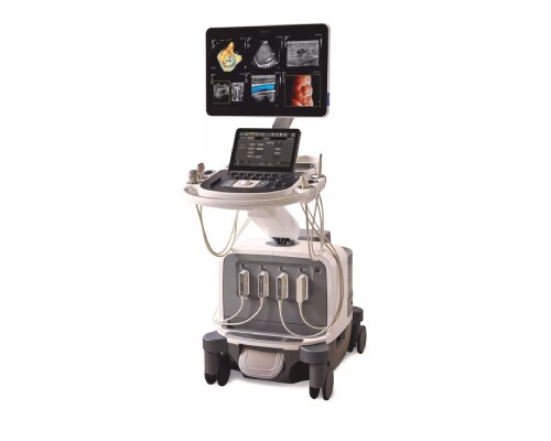
An obstetric ultrasound was conducted at our practice utilizing a Philips EPIQ Elite ultrasound system with the C5-1 curvilinear, eL18-4 linear and V9-2 PureWave volume transducers. The ultrasound evaluation demonstrated a breech female fetus with a normal estimated fetal weight. The fetal anatomic survey did not demonstrate any fetal abnormalities. The placenta was implanted along the anterior uterine wall with no evidence of previa. A suspected placental abnormality was noted along the superior portion of the anterior uterine wall using the C5-1 transducer (Figure 1). The V9-2 volume transducer was used to obtain additional 2D images (Figures 2 and 3).


How the diagnosis was made
The patient was enrolled in a clinical evaluation study of the eL18-4 linear and V9-2 PureWave volume transducers which was ongoing at the time of her consultation at our practice. Utilizing the eL18-4 linear and V9-2 PureWave volume transducers on the Philips EPIQ Elite ultrasound system, additional imaging was performed with particular focus on the suspected placental abnormality and the placental-uterine interface. MicroFlow Imaging (MFI) and MicroFlow Imaging High Definition (MFI HD), designed to enhance the visualization and image resolution of low velocity blood flow, were utilized to assess the placental and uterine anatomy.
Initial 2D imaging using the C5-1 transducer demonstrated a placental abnormality. The findings, based on evaluation solely with the C5-1 transducer, were alarming for a uterine defect due to a placenta percreta. With the use of the V9-2 and eL18-4 transducers, additional images were acquired, better delineating the differences in echotexture between the placenta, uterine myometrium and the rectus abdominis muscles. These images demonstrated the evidence necessary to support the diagnosis of a maternal rectus abdominis separation with the uterine wall bulging into this defect (Figures 4 and 5).


MFI and MFI HD were applied to the placental-uterine interface (Figures 6 and 7). Using the higher frequency V9-2 PureWave volume and eL18-4 PureWave linear transducers, the ultrasound abnormality was explored in greater detail. Through the use of multiple transducers and MFI HD and MFI, the imaging substantiated the diagnosis of a normal placental-uterine interface, no evidence of PAS, but instead, a defect of the rectus abdominis muscles in the patient. She was informed of the finding of a rectus abdominis diastasis (RD) and the good prognosis of this diagnosis as she prepared for her delivery. At 39 0/7 weeks gestation, she underwent a repeat cesarean section, delivering a female infant weighing 7 lbs 5 oz. The placenta was delivered without difficulty with no evidence of PAS.


Summary
Ultrasound continues to be an outstanding, low-cost diagnostic tool and remains the primary imaging modality for the diagnosis of fetal and placental anomalies. Over the last several years, significant advancements in ultrasound continue to change the way clinicians evaluate and diagnose fetal and placental abnormalities. With cutting-edge transducer technology such as the V9-2 PureWave volume transducer and the eL18-4 PureWave linear transducer, our ability to evaluate complex anatomic issues is improving along with a variety of obstetric diagnoses.
In this patient’s case, the suspected PAS diagnosis was not accurate with the use of 2D imaging from the C5-1 transducer alone. The diagnosis of placenta percreta was incorrect because the placental-uterine interface was not well visualized on initial imaging. The higher frequency of the V9-2 PureWave volume transducer and the eL18-4 PureWave linear transducer allowed for greater resolution imaging, while MFI and MFI HDI enhanced the ability to delineate the discrete levels of the placenta, uterine myometrium and rectus abdominis with color vascular flow. A defect ultimately was identified at the level of the maternal rectus abdominis which is correctly characterized as an RD.
As cesarean deliveries increase, the complication rates of PAS rise, along with unique findings such as RD.3 With state-of-the-art tools, including the V9-2 PureWave volume transducer and eL18-4 PureWave linear transducer with MFI and MFI HD, the clinician’s ability to elucidate more accurate diagnoses is enhanced greatly. As in this case, the true diagnosis was made with greater confidence with the use of advanced ultrasound transducers and innovative color Doppler technology.

Michael S. Ruma, MD, MPH
Maternal-Fetal Medicine
Perinatal Associates of New Mexico
Albuquerque, New Mexico, USA
Clinical article
High-frequency transducers and MFI HD enhance the diagnosis of a suspected placental abnormality
Subscribe to our email updates
Subscribe to our email updates
Footnotes
[1] Silver R, Branch DW. Placenta Accreta Spectrum. NEJM. 2018;378:1529–36. [2] Berkley EM, Abuhamad AZ. Prenatal diagnosis of placenta accrete: is sonography all we need? J Ultrasound Med. 2013;32:1345–50. [3] Bakken Sperstad J, Kolberg Tenfjord M, Hilde G, Ellstrom-Eng M, Bo K. Diastasis recti abdominis during pregnancy and 12 months after childbirth: prevalence, risk factors and report of lumbopelvic pain. J Sports Med. 2016 Sep;50(17):1092–6.
Results from case studies are not predictive of results in other cases. Results in other cases may vary.


