FlexVue with Orthogonal View enhances the diagnosis of a fetal facial cleft abnormality
By Michael S. Ruma, MD, MPH ∙ Mar 31, 2020 ∙ 2 min read
Cleft lip, with or without cleft palate, is the most common congenital facial malformation.1 Today, the antenatal diagnosis of a fetus with a cleft lip is commonly made prior to birth with the use of prenatal diagnostic ultrasound. Traditionally, 2D ultrasound stands as the mainstay of fetal evaluation, and the prenatal diagnosis of cleft lip is readily made with this ultrasound mode.
At-a-glance

Nevertheless, with advances in technology, the use of 3D and 4D ultrasound has enhanced not only the diagnosis of cleft lip but also the understanding of the diagnosis by the patient. While cleft lip is commonly noted on 2D ultrasound, the prenatal diagnosis of a cleft palate remains challenging. Differentiating the diagnosis of cleft lip, with or without a palatal defect, is important for the clinicians involved with counseling the parents.
An isolated cleft lip routinely is surgically repaired in a single procedure, while numerous interventions and additional complications may occur with defects of the palate.1
A clinical example: assessing a suspected facial abnormality
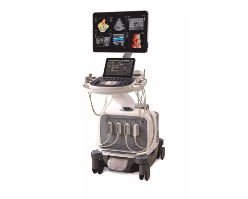
A 34-year-old gravida 1 para 0 pregnant patient at 26 0/7 weeks gestation presented to Perinatal Associates of New Mexico (PANM) for evaluation of a suspected facial abnormality on an outside ultrasound. An obstetric ultrasound was conducted at our practice, utilizing a Philips EPIQ Elite ultrasound system with both the C5-1 curved array transducer and the V9-2 PureWave mechanical volume transducer.
The ultrasound evaluation demonstrated a vertex, female fetus with a normal estimated fetal weight. The fetal anatomic survey identified a unilateral left cleft lip with no additional fetal abnormalities (Figure 1). The Philips V9-2 PureWave volume transducer was used to obtain a 3D volume to provide a surface-rendered image for patient counseling (Figure 2).


How the diagnosis was made
The patient was enrolled in a clinical evaluation study of the Philips V9-2 PureWave volume transducer which was ongoing at the time of her consultation at our practice. Utilizing the Philips V9-2 PureWave volume transducer on the Philips EPIQ Elite ultrasound system, additional imaging was performed with particular focus on the fetal palatal anatomy. The FlexVue with Orthogonal View feature, designed to simplify the evaluation of a 3D volume, was utilized to assess the fetal cleft lip and to evaluate the fetal palate.
2D imaging using the C5-1 and V9-2 transducers provided evidence to support the diagnosis of a unilateral left cleft lip; however, imaging of the fetal palate was limited throughout the ultrasound exam. Visualization of the fetal cleft lip and assessment of the fetal palate were evaluated with the use of the capabilities of FlexVue with Orthogonal View.
First, a 3D volume was obtained using the V9-2 transducer (Figure 3).

Next, the fetal cleft lip was identified by applying the FlexVue line to a sagittal image of the fetal face, demonstrating the corresponding orthogonal (coronal) plane (Figure 4).

Last, the Orthogonal View line was applied demonstrating the corresponding orthogonal (transverse) plane, allowing for interrogation of the fetal palate and demonstration of normal palatal anatomy (Figures 5 and 6).


Manipulation of the 3D volume with FlexVue with Orthogonal View provided additional imaging to substantiate normal anatomy of the fetal palate. The patient was informed of the findings of an isolated unilateral left cleft lip and referred to pediatric plastic surgery for antenatal consultation with a plan for postnatal surgical repair.
Summary
The importance of ultrasound cannot be understated in its place for the prenatal diagnosis of fetal anomalies. Over the last several decades, advances in ultrasound technology have vastly changed the process of making, and the confidence behind, the diagnosis of many fetal abnormalities.
With innovative transducer technology such as the V9-PureWave volume transducer, our reliance on 2D imaging to evaluate fetal anatomy is waning as clinicians at PANM become more confident in utilizing 3D volume rendering. In this patient’s case, the diagnosis was refined more accurately using not only 2D, but also 3D imaging along with cutting-edge tools for interpretation of the fetal 3D volume. With FlexVue with Orthogonal View, the diagnosis was enhanced, providing both the clinician and the patient a higher degree of confidence in the true diagnosis and prognosis of an isolated unilateral cleft lip in the fetus.

Michael S. Ruma
Maternal-Fetal Medicine
Perinatal Associates of New Mexico
Albuquerque, New Mexico, USA
Clinical article
FlexVue with Orthogonal View enhances the diagnosis of a fetal facial cleft abnormality
Subscribe to our email updates
Footnotes
[1] James JN, Schlieder DW. Prenatal Counseling, Ultrasound Diagnosis, and the Role of Maternal-Fetal Medicine of the Cleft Lip and Palate Patient. Oral Maxillofac Surg Clin North Am. 2016;28:145-51.
Results from case studies are not predictive of results in other cases. Results in other cases may vary.


